Arteries In Neck / Department Of Surgery Carotid Artery Disease. Your doctor may then test your physical and mental capabilities such as strength, memory and speech. The vertebral arteries are branches of the subclavian (upper extremity) arteries. If one of them is narrowed or blocked, it can lead to a stroke. Your carotid arteries are two major blood vessels on either side of your neck that provide a critical blood supply to the brain and head. Two large arteries flow from the heart up the sides of the neck and into the brain.
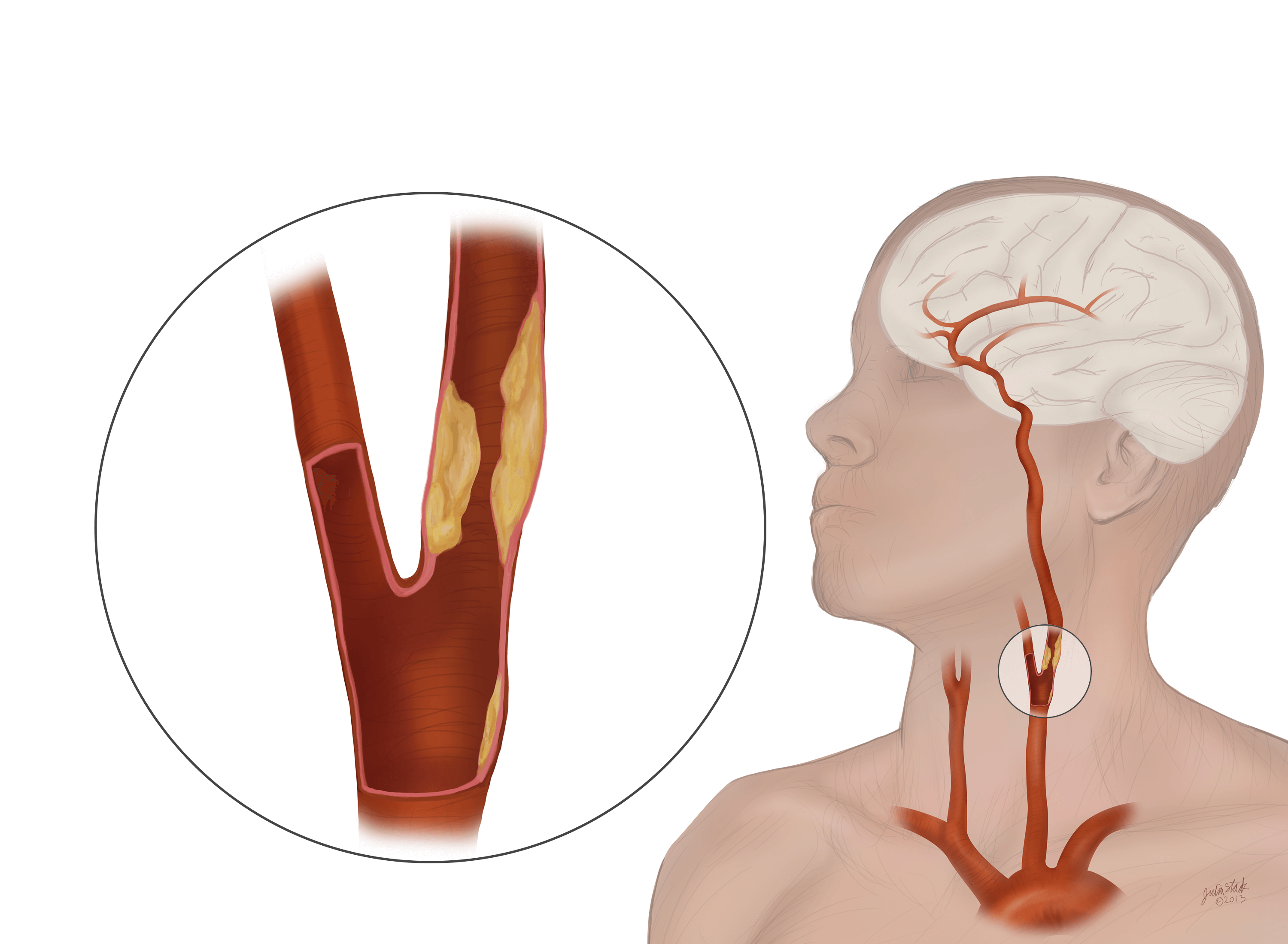
When fatty deposits develop in the area, it can put you at a heightened risk for a blood clot that can disrupt blood flow to your brain. Two large arteries flow from the heart up the sides of the neck and into the brain. In the neck, the carotid sheath (fibrous connective tissue) covers the common carotid artery, vagus nerve, and internal jugular vein. The arteries in neck that supply blood to the brain are called carotid arteries. At the root of the neck the right internal jugular vein is placed at a little distance from the common carotid artery, and crosses the first part of the subclavian artery, while the left internal jugular vein usually overlaps the common carotid artery.
Carotid Artery Disease Johns Hopkins Medicine from www.hopkinsmedicine.org Aches and pains in the jaw and neck are common symptoms of angina, which is the discomfort that results from poor blood flow to part of the heart. These arteries originate from different arteries but follow symmetrical courses. Your doctor may then test your physical and mental capabilities such as strength, memory and speech. Through their course, they give off several meningeal, muscular and spinal branches for the nearby structures. The vein is the most lateral structure within the carotid sheath, followed by the nerve and then the artery, which is the most medial structure. The exam generally includes listening for a swooshing sound (bruit) over the carotid artery in your neck, a sound that's characteristic of a narrowed artery. The left from the aortic arch in the thorax. One carotid artery is located on each side of your neck.
The plaque buildup is made of fat, cholesterol, cellular waste, calcium, proteins and inflammatory cells.
The left from the aortic arch in the thorax. The blockage increases your risk of stroke, a medical emergency that occurs when the blood supply to the brain is interrupted or seriously reduced. A new classification system divides the internal carotid artery into four parts; Your carotid arteries are the major blood vessels that deliver blood to your brain. To continue reading this article, you must log in. Cervical in the neck, petrous in the base of the skull, cavernous within the cavernous sinus and intracranial above the cavernous sinus. Your doctor may then test your physical and mental capabilities such as strength, memory and speech. The right common carotid originates in the neck from the brachiocephalic trunk; The common carotid arteries are present on the left and right sides of the body. The carotid arteries can be felt on each side of the lower neck, immediately below the angle of the jaw. The carotid arteries provide the head's blood supply and run along both sides of the neck. Stroke deprives your brain of oxygen. The left and right common carotid arteries ascend up the neck, lateral to the trachea and the oesophagus.
After that, your doctor may recommend: The arteries in the chest, neck and brain are the most frequent arteries found to be abnormal in phace syndrome. The carotid arteries are two large blood vessels that supply oxygenated blood to the large, front part of the brain. They are the carotid arteries, and they carry blood to the brain. Cadaveric angiographic and dissection studies have demonstrated that the external and internal carotids are the main arterial sources for the head and…
Cardiovascular System Of The Head And Neck from innerbody.imgix.net The pain may be sudden and severe—people often describe it as a throbbing pain. Two pairs of blood vessels in the neck — the carotid and vertebral arteries, known collectively as the cervical arteries — carry blood to the brain. Carotid artery disease occurs when fatty deposits (plaques) clog the blood vessels that deliver blood to your brain and head (carotid arteries). Cervical in the neck, petrous in the base of the skull, cavernous within the cavernous sinus and intracranial above the cavernous sinus. Your carotid arteries are the major blood vessels that deliver blood to your brain. They arise, one on each side of the body, go through the vertebral column (spine) in the back of the neck, and enter the skull via the hole at the base of the skull called the foramen magnum. There are two carotid arteries (one on each side of the neck) that supply blood to the brain. The exam generally includes listening for a swooshing sound (bruit) over the carotid artery in your neck, a sound that's characteristic of a narrowed artery.
Blood leaks between the layers of the artery wall and forms a clot.
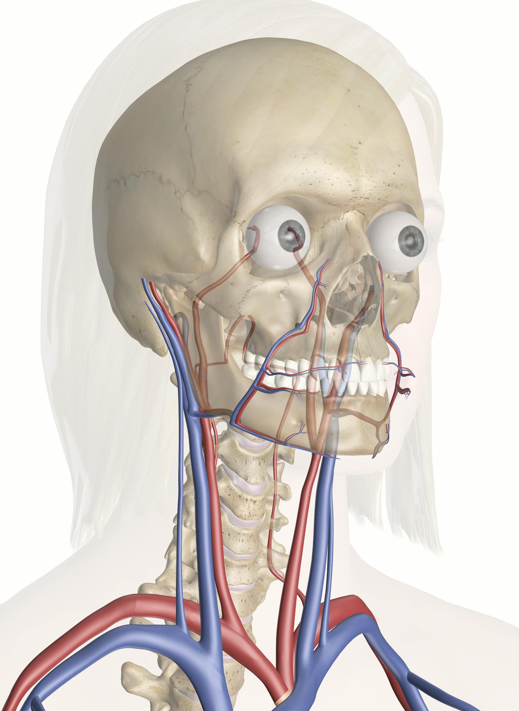
These arteries originate from different arteries but follow symmetrical courses. Stroke deprives your brain of oxygen. Aches and pains in the jaw and neck are common symptoms of angina, which is the discomfort that results from poor blood flow to part of the heart. When fatty deposits develop in the area, it can put you at a heightened risk for a blood clot that can disrupt blood flow to your brain. Veins and arteries of the neck 9 photos of the veins and arteries of the neck activate javascript arteries in the neck diagram, common carotid artery branches, external carotid artery function, how many carotid arteries, left common carotid artery function, the left common carotid artery supplies blood to the. One carotid artery is located on each side of your neck. Doctors can test for a narrowed carotid artery, but it's usually not a good idea. The left from the aortic arch in the thorax. Carotid artery disease occurs when fatty deposits (plaques) clog the blood vessels that deliver blood to your brain and head (carotid arteries). Without this blood flow, your brain cells would. Related posts of arteries in the neck picture veins and arteries of the neck. At the level of the superior margin of the thyroid cartilage (c4), the carotid arteries split into the external and internal carotid arteries. The right common carotid originates in the neck from the brachiocephalic trunk;
Two pairs of blood vessels in the neck — the carotid and vertebral arteries, known collectively as the cervical arteries — carry blood to the brain. They supply oxygen to the parts of the brain that control our movements and our ability to think, speak and experience our senses of touch, taste, sight, sound and feel. However, pain from carotidynia typically only occurs on one side. The blockage increases your risk of stroke, a medical emergency that occurs when the blood supply to the brain is interrupted or seriously reduced. The vein is the most lateral structure within the carotid sheath, followed by the nerve and then the artery, which is the most medial structure.
Carotid Artery Disease Johns Hopkins Medicine from www.hopkinsmedicine.org If one of them is narrowed or blocked, it can lead to a stroke. Two pairs of blood vessels in the neck — the carotid and vertebral arteries, known collectively as the cervical arteries — carry blood to the brain. Veins and arteries of the neck 9 photos of the veins and arteries of the neck activate javascript arteries in the neck diagram, common carotid artery branches, external carotid artery function, how many carotid arteries, left common carotid artery function, the left common carotid artery supplies blood to the. They arise, one on each side of the body, go through the vertebral column (spine) in the back of the neck, and enter the skull via the hole at the base of the skull called the foramen magnum. The carotid arteries can be felt on each side of the lower neck, immediately below the angle of the jaw. When fatty deposits develop in the area, it can put you at a heightened risk for a blood clot that can disrupt blood flow to your brain. Stroke deprives your brain of oxygen. Your carotid arteries are the major blood vessels that deliver blood to your brain.
When your doctor puts their hands on your neck to detect.
At the level of the superior margin of the thyroid cartilage (c4), the carotid arteries split into the external and internal carotid arteries. These arteries originate from different arteries but follow symmetrical courses. After that, your doctor may recommend: Doctors can test for a narrowed carotid artery, but it's usually not a good idea. The carotid arteries provide the head's blood supply and run along both sides of the neck. The arteries in the chest, neck and brain are the most frequent arteries found to be abnormal in phace syndrome. Your carotid arteries are the major blood vessels that deliver blood to your brain. If one of them is narrowed or blocked, it can lead to a stroke. They do not give off any branches in the neck. The carotid arteries are two large blood vessels that supply oxygenated blood to the large, front part of the brain. What are the arteries of the chest, neck and brain? The common carotid arteries are present on the left and right sides of the body. Cadaveric angiographic and dissection studies have demonstrated that the external and internal carotids are the main arterial sources for the head and…
Share :
Post a Comment
for "Arteries In Neck / Department Of Surgery Carotid Artery Disease"

Post a Comment for "Arteries In Neck / Department Of Surgery Carotid Artery Disease"